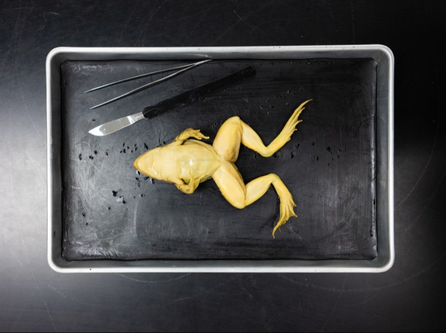
Locate the cloaca at the specimen’s posterior end.In a living frog, this membrane is clear. This is the frog’s third eyelid, the nictitating membrane. Notice the cloudy eyelid attached at the bottom of each eye. Posterior to the eyes are round tympanic membranes, the frog’s external sound receptors.Find the 2 external nares at the head’s tip.Each hind limb is divided into a thigh, lower leg, and foot. Observe that each forelimb is divided into an upper arm, forearm, and hand.


The frog is a tetrapod, meaning that it possesses 4 limbs for locomotion.

In virtual dissection all the frogs are the same. Dissection is a good way to learn about the structure of all living creatures, and it's interesting to see how the frog of another student may be different than yours. In virtual dissection there are no surprises. No student should be forced to partake in actual dissection if he or she objects to it, but the act of making that decision is also an important part of education. If a student has moral objections to frog dissection, that can be a good thing and lead to possibilities for self-discovery. Textbook and Internet images are something students see everyday, but an actual once-living thing stays with a person. Virtual dissection does not create the same lasting impression or vivid memory as actual dissection. It also gives a student the chance to discover whether he or she possesses aptitude for this kind of work. Practice with actual dissection provides good training for such tasks and a good way for a young person to understand the process. Future doctors, biologists, and other scientists may need at some point to perform surgery or dissections.


 0 kommentar(er)
